X-ray Diagnostics
Magnetic resonance imaging (MRI, nuclear magnetic resonance imaging)
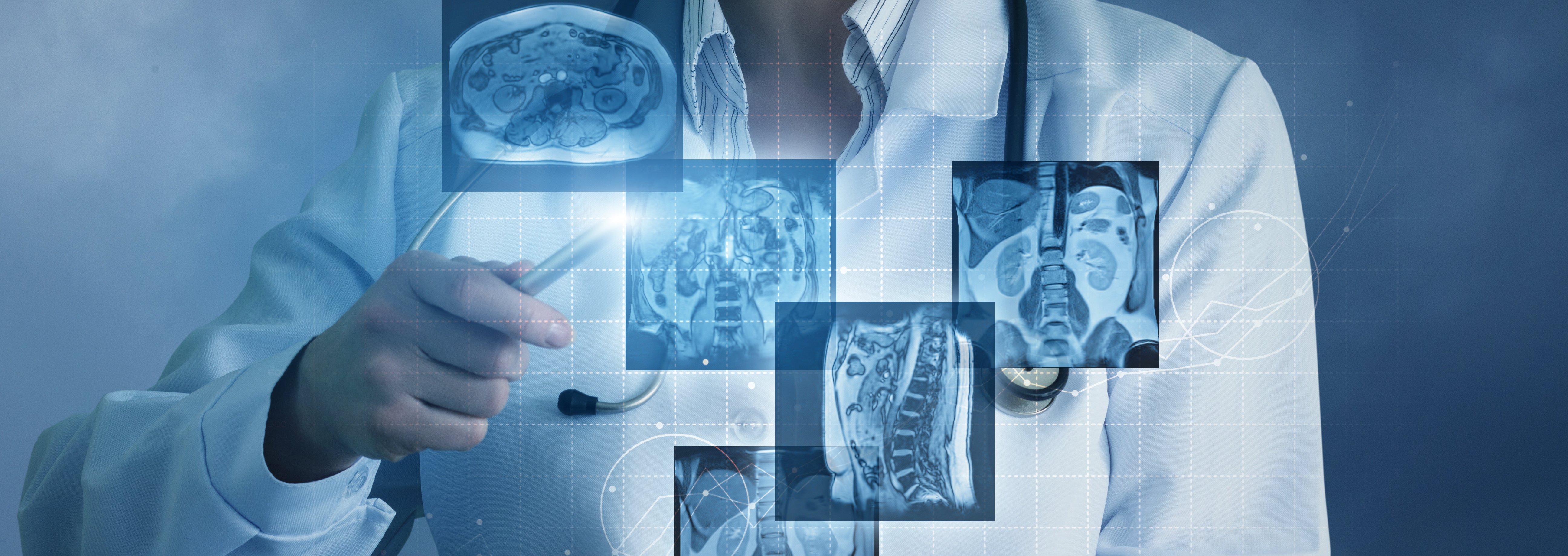
Magnetic resonance imaging (MRI) is used to visualise the structure and function of tissues and organs in the body. In contrast to CT, it does not involve the use of X-rays.
Magnetic resonance imaging (MRI) can also be used to visualise blood vessels, a procedure known as MR angiography or MRA.
There are three techniques to choose from for MR angiography (MRA):
- Time-of-flight (TOF)
- Phase contrast analysis (PCA)
- Contrast-enhanced Magnetic Resonance Angiography (ceMRA)
Time-of-flight (TOF) and phase contrast analysis (PCA) make use of artefacts caused by blood flow to produce high-signal images of flowing blood and thus of blood vessels.
Areas of application for these techniques are
- Examination of cerebral arteries,
- Cerebral veins and
- Large veins of the abdomen and pelvis.
Contrast-enhanced ceMRA
Contrast-enhanced ceMRA, which has only recently been established, is becoming increasingly important in clinical practice. A highly tolerable paramagnetic contrast agent that can also be used for patients with renal insufficiency is injected intravenously, thereby increasing the signal levels in blood. This makes this technique less artefact-dependent than the two methods described above. Measurement time is in the range of seconds, around 10 to 30 seconds.
![[Translate to englisch:] Siegel FAZ Deutschlands beste Krankenhäuser](/fileadmin/_processed_/3/8/csm_Siegel_FAZ_Beste_Krankenhaeuser_2026_Universitaetsklinikum_Regensburg_f1e64de630.png)


![[Translate to englisch:] Siegel Beruf & Familie geht gut bei uns](/fileadmin/_processed_/8/e/csm_lkr_siegel_beruf_fam2026_9a13be1ba5.jpg)